seungahlee@snu.ac.kr
Department of Mechanical Engineering, Seoul National University

Computational microscopy: Fourier ptychography, phase and polarization imaging, virtual staining

Seung Ah Lee's Imaging Intelligence Laboratory at Seoul National University develops Fourier ptychographic microscopes (FPM) for wide-field, high-resolution quantitative phase imaging: a smartphone FPM (K. C. Lee et al., ACS Photonics 2021), FPM with an OLED display as the light source (K. Lee et al., Opt. Lasers Eng. 2024), single-shot FPM (H. Yoon et al., Appl. Phys. Lett. 2025) and polarization-resolved FPM (H. Chae et al., Laser Photonics Rev. 2026), also used for virtual staining (H. Chae et al., APL Photonics 2024).

Summary

A conventional microscope trades field of view for resolution, and unstained cells give little contrast. Fourier ptychography (FP) illuminates the sample from many angles and combines the low-resolution images computationally into a wide-field, high-resolution image with quantitative phase. The illumination comes from an LED array or a display screen, and the images are recorded through an inexpensive low-magnification objective. We have built smartphone, single-shot and polarization-resolved FP microscopes.

Code, data and patents

  • Codegithub.com/IIL-SNU/FP_ADMMRegularized (ADMM) Fourier ptychography reconstruction. K. C. Lee et al., Optics Express 2024.
  • Codegithub.com/IIL-SNU/Smartphone_FPMA smartphone-based Fourier ptychographic microscope using the display screen for illumination. K. C. Lee et al., ACS Photonics 2021.
  • DataSmartphone FPM example imagesRaw image stacks captured with the smartphone Fourier ptychographic microscope (display-screen illumination) for a blood smear, a phase target and a USAF resolution target, with the Android app (APK) used to acquire them.
  • PatentUS11880965B2Smartphone for obtaining Fourier ptychography image and method for obtaining Fourier ptychography image using smartphone (granted 2024)
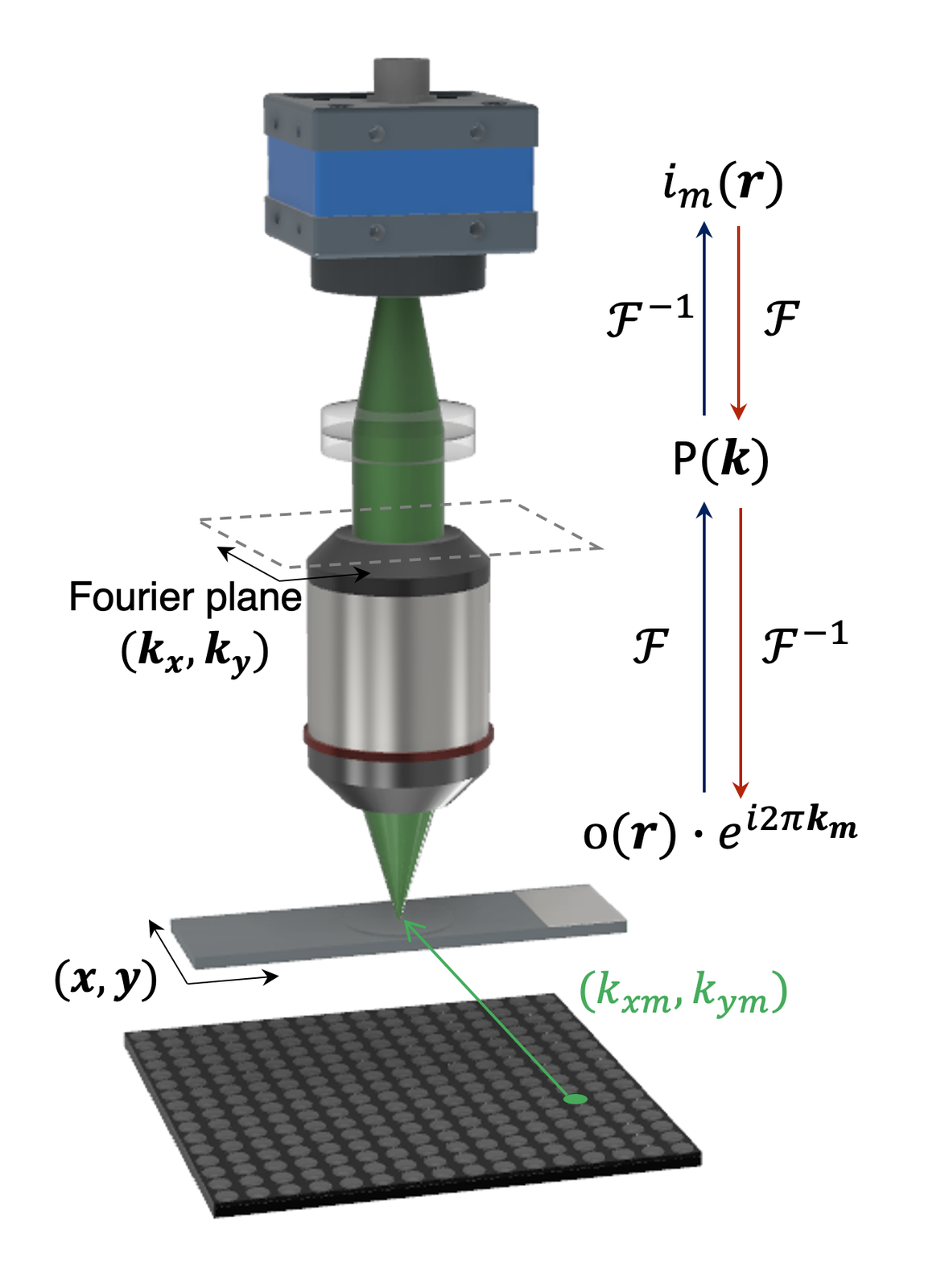
FP forward model: tilted LED illumination shifts the sample spectrum through the objective pupil onto the camera.
FP forward model: tilted LED illumination shifts the sample spectrum through the objective pupil onto the camera. IIL (conference presentation)
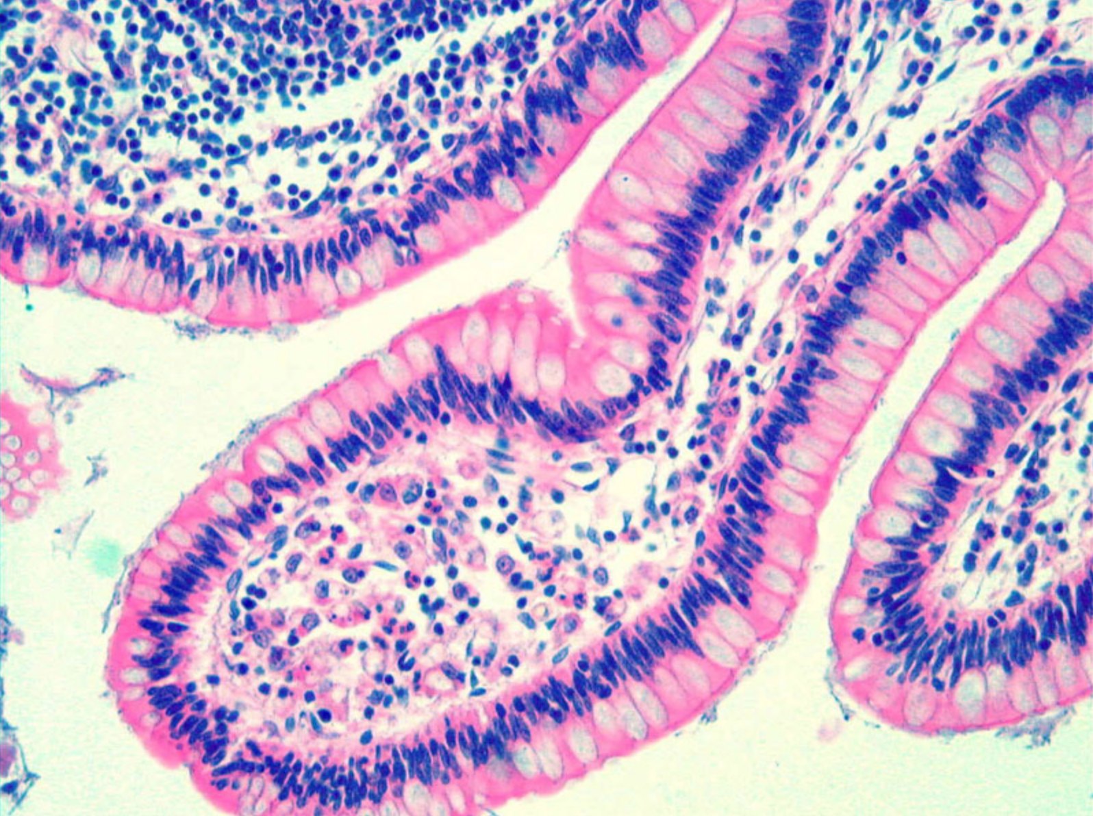
High-resolution FP image of an H&E-stained tissue section recovered with a low-magnification objective.
High-resolution FP image of an H&E-stained tissue section recovered with a low-magnification objective. IIL (conference presentation)

Key results

75 fps
single-shot quantitative phase video of swimming microorganisms Yoon et al., Appl. Phys. Lett. 2025
2.5×
resolution beyond the objective's diffraction limit, from one snapshot Yoon et al., Appl. Phys. Lett. 2025
870 nm
half-pitch resolution over 2.1 × 1.6 mm² on a stand-alone smartphone microscope K. C. Lee et al., ACS Photonics 2021
1 camera
full Jones-matrix birefringence imaging with one monochrome sensor and no moving parts Chae et al., Laser Photonics Rev. 2026

Projects

7 projects

2019–2021

Smartphone Fourier ptychographic microscope

A smartphone has a camera, a programmable display and a processor. We combined them into a stand-alone Fourier ptychographic microscope (FPM). A compact 3D-printed optics module guides light from the phone's own screen through the sample and into its built-in camera, so lighting up individual spots on the display becomes a programmable, angle-scanning illumination source.

View on the research page

2020–2022

Live-cell phase imaging inside the incubator

Live-cell imaging usually trades field of view for resolution and often relies on fluorescent labels that can perturb the cells. We built a miniaturized Fourier ptychographic microscope small enough to operate inside a standard CO₂ cell-culture incubator, using a low-magnification 4× objective, a machine-vision camera and an inexpensive LED matrix.

View on the research page

2021–2024

Display screens as programmable FP illumination

FP is usually illuminated by an LED array, whose millimeter-scale LEDs must sit far from the sample to approximate plane waves. An OLED display instead provides millions of micrometer-sized emitters that can be switched on in any pattern. We studied how to use a high-resolution display screen, placed just beneath the slide, as the illumination source.

View on the research page

2009–2014

Chip-scale lensless microscopy (PhD roots at Caltech)

Before the lab existed, Prof. Lee's PhD work at Caltech (in Changhuei Yang's group) asked how small and inexpensive a microscope could be. In chip-scale microscopy the sample sits directly on a CMOS image sensor and casts a shadow onto its pixels; sequences of slightly shifted shadow images are then combined by pixel super-resolution to beat the pixel-size limit.

View on the research page

Computational Microscopy

Virtual staining

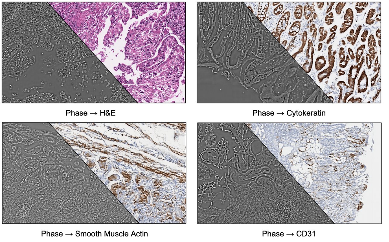
Seung Ah Lee's laboratory develops virtual staining for digital pathology: neural networks convert the quantitative phase of unstained tissue slides, measured by FPM, into images of stained slides (H. Chae et al., APL Photonics 2024; K. C. Lee et al., J. Phys. Photonics 2026).

Summary

Pathologists read tissue through chemical stains such as hematoxylin and eosin (H&E). Staining is slow, consumes tissue and reagents, varies from lab to lab, and fades over the years. Unstained tissue is nearly transparent, but it still delays light by an amount set by its local density and thickness. Our Fourier ptychographic microscopes measure this optical phase quantitatively across whole slides, and deep neural networks learn to translate it into the colors of a stained slide. We use this to restore faded archival slides digitally, to co-design illumination and networks so that a slide region is scanned in seconds, and to explore stains beyond H&E, working with pathologists at Severance Hospital.

Code, data and patents

  • Codegithub.com/IIL-SNU/FPVSFourier ptychographic virtual staining. K. C. Lee et al., J. Phys. Photonics 2026.
  • DataFPVS example FPM dataExample full-field-of-view Fourier ptychographic microscopy inputs and reconstructions for the virtual staining code.
  • DataDigital staining literature databaseTable of all articles covered in the digital staining review with their categorizations, published as the review's supplementary files.
Label-free phase images split diagonally with virtual H&E and virtual immunohistochemistry.
Label-free phase images split diagonally with virtual H&E and virtual immunohistochemistry. IIL (conference presentation)

Key results

~10 yr
old, faded H&E slides from a hospital archive restored by virtual re-staining Chae et al., APL Photonics 2024
16 patterns
faster slide scanning than single-LED FP, using 16 learned illumination patterns instead of 193 single LEDs K. C. Lee et al., J. Phys. Photonics 2026
~5 s
to scan a 4 mm × 3 mm tissue area, with about 20 ms of virtual staining per field of view K. C. Lee et al., J. Phys. Photonics 2026

Virtual staining projects

3 projects

2021–2024

Restoring faded slides with NIR phase-to-color virtual staining

Hematoxylin and eosin stains fade over time, so archived slides that may be needed again for diagnosis or research slowly lose their information. Physical re-staining is laborious and can damage the tissue, and digital color normalization fails once the stain is nearly gone.

View on the research page

2022–2025

High-speed virtual staining with learned illumination

Two factors limit virtual staining in clinical workflows: collecting paired stained and unstained training images is costly, and capturing hundreds of FP images per field is slow. We addressed both with a co-designed illumination and network system. An illumination-optimization layer, trained end to end with the network, learns which combinations of the microscope's 193 LEDs carry the most useful information.

View on the research page

Publications

23 publications

  1. Computational space-bandwidth product expansion via multiscale aperture synthesis and wavefront engineering

    Kang, Y.G., Chae, H., & Lee, S.A.

    Advanced Imaging, 3(3), 033001 (2026) doi:10.3788/AI.2026.30001

  2. Pupil Plane Multiplexing for Vectorial Fourier Ptychography

    Chae, H., Lee, K., Kang, Y.G., Yoon, H., Lee, K.C., & Lee, S.A.

    Laser & Photonics Reviews, 20(9), e02183 (2026) doi:10.1002/lpor.202502183

  3. Semi-supervised virtual staining using learned-illumination Fourier ptychography for high-speed label-free histopathology

    Lee, K.C., Chae, H., Kim, J., Kreiss, L., Kim, H., Kang, Y.G., Zhou, K.C., Chaware, A., Kim, K., Xu, S., Kang, S., Bang, G., Cho, N.H., Hwang, D., Horstmeyer, R., & Lee, S.A.

    Journal of Physics: Photonics, 8(1), 015025 (2026) doi:10.1088/2515-7647/ae270f

  4. Single-shot Fourier ptychography using polarization-encoded illumination

    Yoon, H., Chae, H., Lee, K.C., Lee, K., & Lee, S.A.

    Applied Physics Letters, 126(12), 123702 (2025) doi:10.1063/5.0248101

  5. Restoring H&E stain in faded slides via phase-to-color virtual staining in near-infrared

    Chae, H., Kim, J., Jeon, J., Lee, K., Lee, K.C., Choi, J.U., Kang, S., Choi, S., Bang, G., Lee, J.H., Park, E., Cho, N.H., & Lee, S.A.

    APL Photonics, 9(10), 106112 (2024) doi:10.1063/5.0226277

  6. Anisotropic regularization for sparsely sampled and noise-robust Fourier ptychography

    Lee, K.C., Chae, H., Xu, S., Lee, K., Horstmeyer, R., Lee, S.A., & Hong, B.W.

    Optics Express, 32(14), 25343–25361 (2024) doi:10.1364/OE.529023

  7. High-resolution display screen as programmable illumination for Fourier ptychography

    Lee, K., Lee, K.C., Jung, J., Chae, H., & Lee, S.A.

    Optics and Lasers in Engineering, 177, 108121 (2024) doi:10.1016/j.optlaseng.2024.108121

  8. Computational 3D topographic microscopy from terabytes of data per sample

    Zhou, K.C., Harfouche, M., Zheng, M., Jönsson, J., Lee, K.C., Kim, K., Appel, R., Reamey, P., Doman, T., Saliu, V., Horstmeyer, G., Lee, S.A., & Horstmeyer, R.

    Journal of Big Data, 11(1), 62 (2024) doi:10.1186/s40537-024-00901-0

  9. Tensorial tomographic Fourier ptychography with applications to muscle tissue imaging

    Xu, S., Yang, X., Ritter, P., Dai, X., Lee, K.C., Kreiss, L., Zhou, K.C., Kim, K., Chaware, A., Neff, J., Glass, C., Lee, S.A., Friedrich, O., & Horstmeyer, R.

    Advanced Photonics, 6(2), 026004 (2024) doi:10.1117/1.AP.6.2.026004

  10. Digital staining in optical microscopy using deep learning - a review

    Kreiss, L., Jiang, S., Li, X., Xu, S., Zhou, K.C., Lee, K.C., Mühlberg, A., Kim, K., Chaware, A., Ando, M., Barisoni, L., Lee, S.A., Zheng, G., Lafata, K.J., Friedrich, O., & Horstmeyer, R.

    PhotoniX, 4, 34 (2023) doi:10.1186/s43074-023-00113-4

  11. Noniterative sub-pixel shifting super-resolution lensless digital holography

    Lee, H., Kim, J., Kim, J., Jeon, P., Lee, S.A., & Kim, D.

    Optics Express, 29(19), 29996–30006 (2021) doi:10.1364/OE.433719

  12. A Smartphone-Based Fourier Ptychographic Microscope Using the Display Screen for Illumination

    Lee, K.C., Lee, K., Jung, J., Lee, S.H., Kim, D., & Lee, S.A.

    ACS Photonics, 8(5), 1307–1315 (2021) doi:10.1021/acsphotonics.1c00350

  13. Disordered Nanocomposite Islands for Nanospeckle Illumination Microscopy in Wide‐Field Super‐Resolution Imaging

    Yoo, H., Lee, H., Rhee, W.J., Moon, G., Lee, C., Lee, S.A., Shin, J.S., & Kim, D.

    Advanced Optical Materials, 9(15), 2100211 (2021) doi:10.1002/adom.202100211

  14. Surface Plasmon Localization-Based Super-resolved Raman Microscopy

    Lee, H., Kang, K., Mochizuki, K., Lee, C., Toh, K.A., Lee, S.A., Fujita, K., & Kim, D.

    Nano Letters, 20(12), 8951–8958 (2020) doi:10.1021/acs.nanolett.0c04219

  15. LudusScope: accessible interactive smartphone microscopy for life-science education

    Kim, H., Gerber, L.C., Chiu, D., Lee, S.A., Cira, N.J., Xia, S.Y., & Riedel-Kruse, I.H.

    PLOS ONE, 11(10), e0162602 (2016) doi:10.1371/journal.pone.0162602 Before IIL

  16. A smartphone-based chip-scale microscope using ambient illumination

    Lee, S.A., & Yang, C.

    Lab on a Chip, 14(16), 3056–3063 (2014) doi:10.1039/C4LC00523F Before IIL

  17. Imaging and identification of waterborne parasites using a chip-scale microscope

    Lee, S.A., Erath, J., Zheng, G., Ou, X., Willems, P., Eichinger, D., Rodriguez, A., & Yang, C.

    PLOS ONE, 9(2), e89712 (2014) doi:10.1371/journal.pone.0089712 Before IIL

  18. Microfluidic-integrated laser-controlled microactuators with on-chip microscopy imaging functionality

    Jung, J.H., Han, C., Lee, S.A., Kim, J., & Yang, C.

    Lab on a Chip, 14(19), 3781–3789 (2014) doi:10.1039/C4LC00790E Before IIL

  19. Chip-scale fluorescence microscope based on a silo-filter complementary metal-oxide semiconductor image sensor

    Lee, S.A., Ou, X., Lee, J.E., & Yang, C.

    Optics Letters, 38(11), 1817–1819 (2013) doi:10.1364/OL.38.001817 Before IIL

  20. On-chip continuous monitoring of motile microorganisms on an ePetri platform

    Lee, S.A., Zheng, G., Mukherjee, N., & Yang, C.

    Lab on a Chip, 12(13), 2385–2390 (2012) doi:10.1039/C2LC40090A Before IIL

  21. Color capable sub-pixel resolving optofluidic microscope and its application to blood cell imaging for malaria diagnosis

    Lee, S.A., Leitao, R., Zheng, G., Yang, S., Rodriguez, A., & Yang, C.

    PLOS ONE, 6(10), e26127 (2011) doi:10.1371/journal.pone.0026127 Before IIL

  22. The ePetri dish, an on-chip cell imaging platform based on subpixel perspective sweeping microscopy (SPSM)

    Zheng, G., Lee, S.A., Antebi, Y., Elowitz, M.B., & Yang, C.

    Proceedings of the National Academy of Sciences, 108(41), 16889–16894 (2011) doi:10.1073/pnas.1110681108 Before IIL

  23. Sub-pixel resolving optofluidic microscope for on-chip cell imaging

    Zheng, G.*, Lee, S.A.*, Yang, S.*, & Yang, C.

    Lab on a Chip, 10(22), 3125–3129 (2010) doi:10.1039/C0LC00213E Before IIL
All research themes All publications