seungahlee@snu.ac.kr
Department of Mechanical Engineering, Seoul National University

Dynamic speckle imaging

Seung Ah Lee's Imaging Intelligence Laboratory at Seoul National University develops rolling-shutter speckle imaging (RSSI), which measures blood flow from a single frame of a standard CMOS sensor (C. Yi et al., Optica 2022; C. Yi et al., Biomed. Opt. Express 2024), with applications to fingertip pulse waveforms (Y. Lee et al., Biomed. Opt. Express 2024) and real-time analysis by physics-guided neural networks (S. Byun et al., J. Phys. Photonics 2026).

Summary

We analyze speckle produced by dynamic scattering to measure the dynamics of a sample. When laser light enters living tissue, moving red blood cells make the speckle pattern fluctuate, at a rate set by the flow speed. Measuring the fluctuation directly usually requires cameras running at more than ten thousand frames per second. Rolling-shutter speckle imaging (RSSI) uses the row-by-row exposure of CMOS sensors, in which successive rows are exposed microseconds apart, so that a single frame records the speckle dynamics. With elongated speckles and a physical model, one frame gives both the flow speed and the fraction of moving scatterers. Applications include cerebral blood-flow mapping, pulse waveforms at the fingertip and real-time analysis with physics-guided neural networks.

Code, data and patents

  • Codegithub.com/IIL-SNU/RSSISingle-shot temporal speckle correlation imaging using rolling shutter image sensors. Yi et al., Optica 2022.
  • DataRSSI in vivo mouse brain speckle imagesRaw single-shot rolling-shutter speckle images of mouse cerebral blood flow (two imaging sessions) used in the RSSI paper, the data cited in its data availability statement.
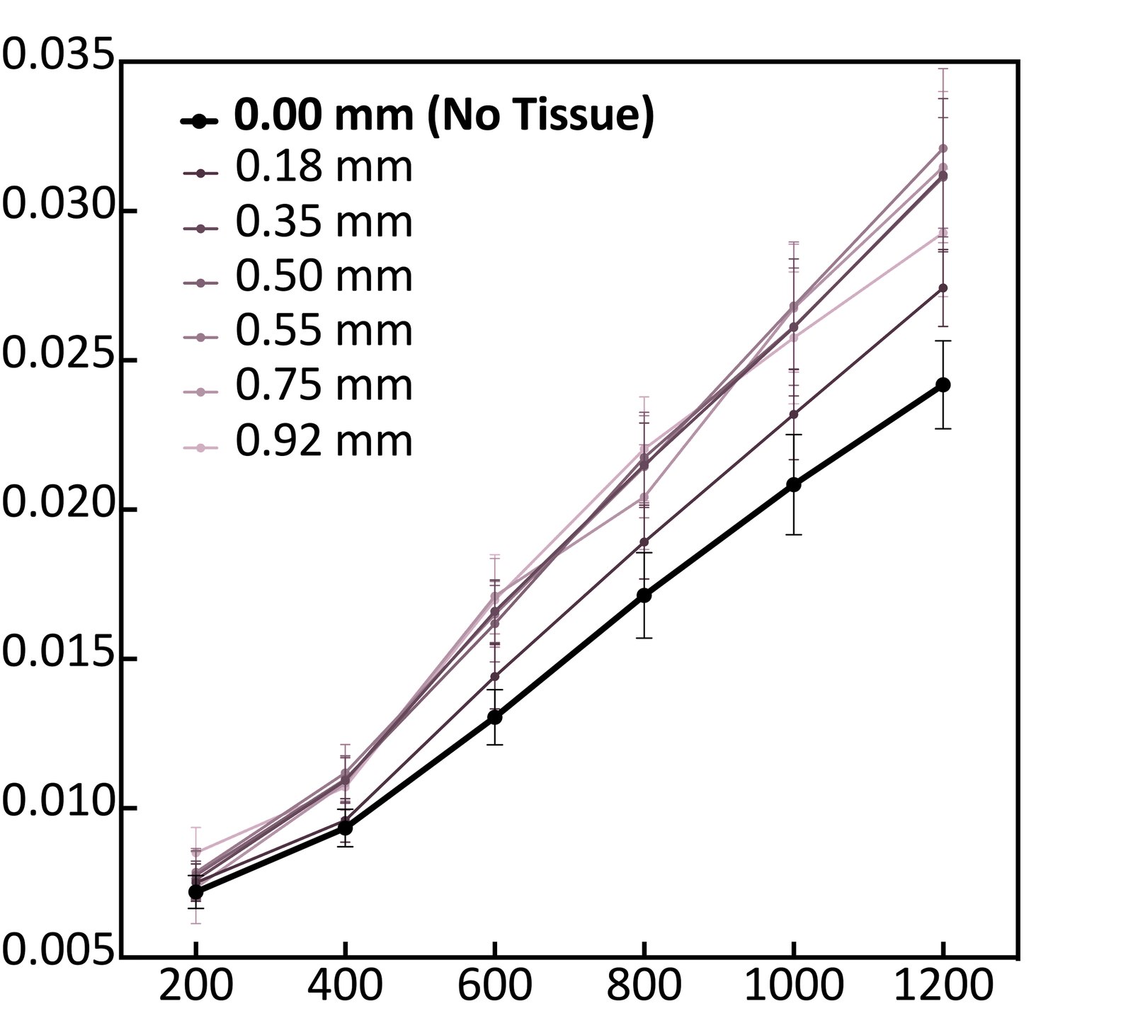
Flow-phantom validation: the measured decorrelation rate rises linearly with flow rate for several tissue-phantom thicknesses (x: flow rate, y: inverse decorrelation time).
Flow-phantom validation: the measured decorrelation rate rises linearly with flow rate for several tissue-phantom thicknesses (x: flow rate, y: inverse decorrelation time). C. Yi et al., Biomed. Opt. Express 2024
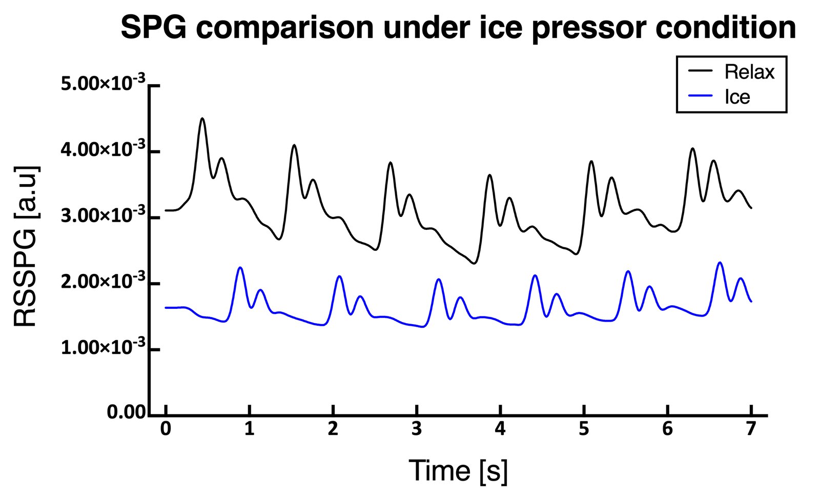
RSSPG during a cold-pressor test compared with relaxation.
RSSPG during a cold-pressor test compared with relaxation. Y. Lee et al., Biomed. Opt. Express 2024

Key results

12 µs
row-to-row delay of a standard rolling-shutter sensor, used as a built-in high-speed clock Yi et al., Biomed. Opt. Express 2024
0.14–2.22 cm/s
phantom flow-speed range validated beneath static tissue layers 0.12–1.56 mm thick Yi et al., Biomed. Opt. Express 2024
14×
more distinct dicrotic notch than photoplethysmography (mean notch ratio 39.3 vs 2.8) Lee et al., Biomed. Opt. Express 2024
4 cm
source–detector separation for deep-tissue blood flow with a 500 × 500 SPAD array Kreiss et al., Neurophotonics 2025

Projects

6 projects

2022–2023

Rolling-shutter speckle imaging (RSSI)

Laser speckle imaging maps blood flow without dyes, but quantitative measurements require following how the speckle decorrelates over microseconds, which normally means costly cameras running above ten thousand frames per second. RSSI instead uses the row-by-row exposure of CMOS sensors, in which successive rows are exposed microseconds apart.

View on the research page

2023–2024

A complete spatiotemporal speckle-correlation model

A single RSSI frame becomes a measurement only once we know how the row-by-row correlation should behave for a given flow, in tissue where moving blood is mixed with static scatterers. Our first model assumed an instantaneous exposure, but real sensors integrate light for longer than the delay between rows.

View on the research page

2023–2024

Rolling-shutter speckle plethysmography (RSSPG)

Photoplethysmography (PPG), the green light on the back of a smartwatch, tracks how much blood lies under the sensor but not how fast it moves. RSSPG applies rolling-shutter speckle imaging to a sequence of frames of a fingertip and extracts, frame by frame, both the decorrelation time, which follows flow speed, and the dynamic scattering fraction, which follows blood volume.

View on the research page

2024–2026

Physics-guided neural networks for real-time speckle imaging

Single-shot RSSI trades statistics for speed: each map is estimated from relatively few speckle grains, so pixel-wise curve fitting is both noisy and slow. Averaging many frames reduces the noise but gives up the time resolution of single-shot imaging.

View on the research page

2025–, ongoing

Speckle-based eye-movement tracking

Fast eye movements, from saccades to the tiny microsaccades made while fixating, carry information about attention and neurological health, yet capturing them usually calls for high-speed cameras. We are extending rolling-shutter speckle imaging to the eye: laser light scattered from the white of the eye (the sclera) forms elongated speckles on a rolling-shutter sensor.

  • S. Byun et al., “Rapid eye movement detection via rolling shutter speckle imaging,” SPIE-ABC 2025, Jeju (poster)

View on the research page

2025

Deep-tissue blood flow with massively parallel DCS

Speckle fluctuations also reveal blood flow far below the surface. Diffuse correlation spectroscopy (DCS) measures how laser light flickers after diffusing through centimeters of tissue, but in adults the light that reaches the brain, beneath scalp and skull, returns faint and noisy.

View on the research page

Publications

5 publications

  1. Physics-guided neural network for single-shot real-time dynamic speckle imaging

    Byun, S., Yi, C., Bae, D., Kang, Y.G., Kim, J., Chang, H., & Lee, S.A.

    Journal of Physics: Photonics, 8(4), 045002 (2026) doi:10.1088/2515-7647/ae99ba

  2. Beneath the surface: revealing deep-tissue blood flow in human subjects with massively parallelized diffuse correlation spectroscopy

    Kreiss, L., Wu, M., Wayne, M., Xu, S., McKee, P., Dwamena, D., Kim, K., Lee, K.C., Cowdrick, K.R., Liu, W., Ülkü, A., Harfouche, M., Yang, X., Cook, C., Lee, S.A., Buckley, E., Bruschini, C., Charbon, E., Huettel, S., & Horstmeyer, R.

    Neurophotonics, 12(2), 025007 (2025) doi:10.1117/1.NPh.12.2.025007

  3. Rolling shutter speckle plethysmography for quantitative cardiovascular monitoring

    Lee, Y., Byun, S., Yi, C., Jung, J., & Lee, S.A.

    Biomedical Optics Express, 15(3), 1540–1552 (2024) doi:10.1364/BOE.511755

  4. Improvements and validation of a spatiotemporal speckle correlation model for rolling shutter speckle imaging

    Yi, C., Byun, S., Lee, Y., & Lee, S.A.

    Biomedical Optics Express, 15(2), 1253–1267 (2024) doi:10.1364/BOE.514497

  5. Single-shot temporal speckle correlation imaging using rolling shutter image sensors

    Yi, C., Jung, J., Im, J., Lee, K.C., Chung, E., & Lee, S.A.

    Optica, 9(11), 1227–1237 (2022) doi:10.1364/OPTICA.465361

All research themes All publications